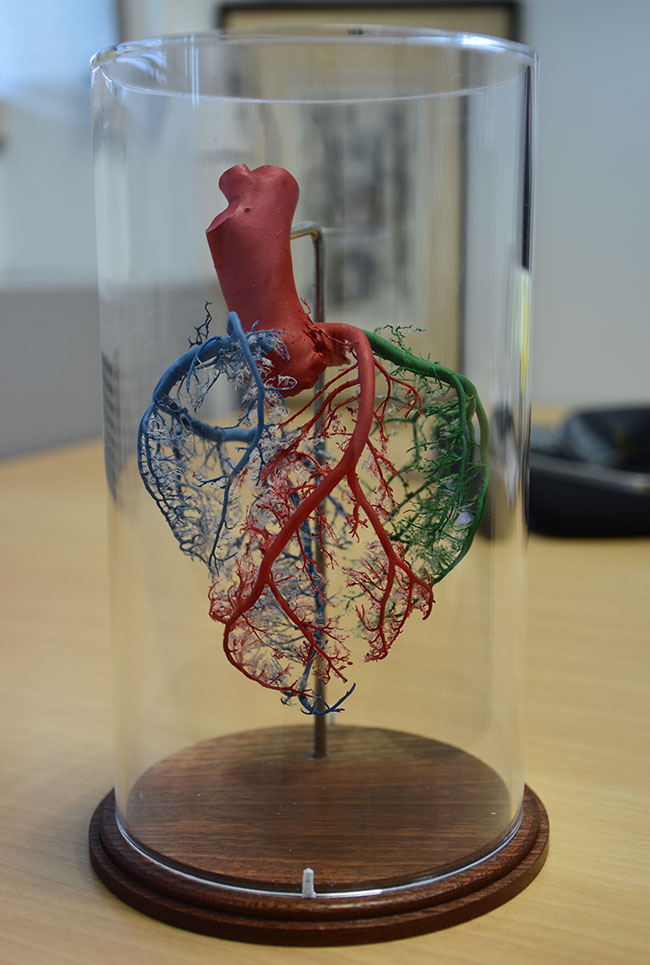
On a shelf in Professor Vicky Cameron's office is something that looks like a piece of art, housed in a glass cylinder to protect it. The heart, striking in its intricate detail, is in fact made of coloured resin.
It is the creation of Professor Greg Jones of the Department of Surgical Sciences at the University of Otago, who works closely with the Christchurch Heart Institute's Professor Cameron, and who uses these models to teach medical students about anatomy.
Professor Jones casts the models by using donated body parts that members of the public have donated for use in medical research and teaching.

The model in Professor Vicky Cameron's office.
Different coloured resin is poured into the blood vessels or tissue of the body part, which may be a heart, kidney, brain, liver or even a hand.
“The idea for this evolved from some research I did many years ago, using it as a way to identify structural defects in blood vessels. The basic technique itself is very old though, having been used to prepare teaching models for anatomy museums for over a 100 years,” Professor Jones said.
Whether the model is to be used for research analysis or teaching, determines the type of resin used. Research requires an expensive resin that allows fine detail to be seen, such as the impression of a cell. Cheaper resin is used in teaching aids.
The models vary in their composition depending on what they are being used to teach.
“They may be used to interpret an angiogram (an x-ray of blood vessels), to understand where heart bloods vessels are lying with a 3D view, the normal variations in blood supply to the heart that occur between individuals and for teaching about surgical procedures such as heart bypass or valve repair.”
More senior doctors such as Registrars are finding the models useful in learning accuracy of needle insertion near the coronary arteries.
Professor Jones gives away his models to teaching Professors and has gifted several sets to the Anatomy Museum at the Otago Medical School.