About the setup
 Cathodoluminescence microscopy equipment in the Department of Geology
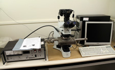
The system we have is a cold cathode system. There are a few parts to this equipment:
- Main equipment
- We have an Olympus BX41 microscope with a trinoc head
- The stage is a technosyn Cold cathode stage
- The control unit controls a standard vacuum pump, the gun and current
- Image capture part
- We have a custom mount and a Canon 1100D digital Single Lens Reflex (dSLR) camera that are attached to the third port on the trinoc head of the microscope
- There is a computer that is used to control the camera, store the saved images and add scale bars etc.
Sample Images
Here are some sample images with captions supplied by Alex Wilson.

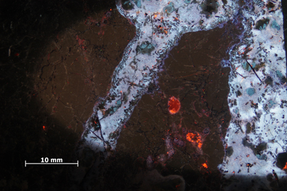
CL image taken at 1 minute exposure time. The CL image is of the contact between a polycrystalline quartz xenolith (right) and tholeitic basalt (left) from the Tawhiroko Sill intrusion in Moeraki Peninsula, NZ. Basalt phases of plagioclase and feldspar-rich, microcrystalline glass around the contact fluoresce with a bright blue, whilst the quartz shows a dull brown. The brightly luminescing gold mineral in the centre is a large grain of apatite.

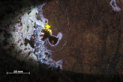
CL image taken at a 30 second exposure time. This CL image is of an injection veinlett of tholeiitic basaltc glass into a polycrystalline quartz xenolith. The contact around the edges of the vein (centre-bright blue), show significant growth of small, lowly-luminescent augite, a product of the addition of silica (quartz xenolith) into the basaltic system. Local silica saturation has occured, indicated by the bright blue, angular expression of recrystallised quartz within the basalt. Carbonate filled cavities produce a strong flourescence of bright orange-red.