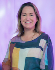
 2018 Chaffer Fellow, Dr Kelly Rogers, will deliver lectures on advanced microscopy techniques, a research seminar about her latest work, and a workshop on image analysis to staff and students at the University of Otago.
Dr Rogers is the Laboratory Head, Centre for Dynamic Imaging Walter and Eliza Hall Institute of Medical Research (WEHI), Melbourne, Australia. She will deliver two lectures on advanced fluorescence microscopy techniques, a workshop on image analysis and present a research seminar on High resolution 4-dimensional microscopy for gaining new insights into mechanisms causing disease.
Her research seminar is on her recent work where live cell super-resolution microscopy using 3D SIM has been used in parallel with Lattice light sheet microscopy to understand critical processes involved in the release of mitochondrial DNA during apoptosis and red blood cell infection by the malaria parasite. The lectures, seminar, and workshop will be available for researchers and postgraduate students from across the University.
Dr Rogers is one of two 2018 Chaffer Fellows. This fellowship supports visits to Otago Medical School by distinguished overseas scientists or clinicians.
Chaffer Fellowship events
1 October 2018, 2–4 pm, Hercus D'Ath Lecture Room
Lecture: Introduction to Fluorescence Microscopy
Part 1: Fluorescence Microscopy Basics
Part 2: State-of-the-Art in Fluorescence Microscopy
For more information and bookings email laura.gumy@otago.ac.nz
2 October 2018, 9am–12 pm, Hercus CAL Lab
Workshop: Image analysis with IMARIS and ImageJ/FIJI
Limited to 50 participants, sign-up deadline: 28 September
For more information and bookings email laura.gumy@otago.ac.nz
2 October 2018, 2pm Barnett Lecture Theatre Dunedin Hospital
Research seminar: High resolution 4-dimensional microscopy for gaining
new insights into mechanisms causing disease
3 October 2018, 2–3 pm, Hercus D'Ath Lecture Room
Lecture: Advanced Techniques in Live Cell Imaging
For more information and bookings email laura.gumy@otago.ac.nz
More about Dr Kelly Rogers
Dr Rogers has 20 years' experience in optical microscopy, particularly in Ca2+ imaging and live cell imaging techniques. She completed her PhD at Griffith University in the Genomics Research Centre in 2001 and then spent 7 years as a postdoctoral fellow at the Institut Pasteur in Paris. While at Pasteur, she developed novel Ca2+ imaging methods to study neural connectivity in the developing mouse brain. In 2009 she joined WEHI where she established the Centre for Dynamic Imaging. Then in 2017 was appointed Laboratory Head as an extension of this role to develop quantitative microscopy. Her team has built a Lattice light sheet microscope and are early adopters of this technology.