Managing some of the resources generated from body donors has become more challenging with the rise of digital technology, says Otago Medical School Senior Lecturer Dr Jon Cornwall.

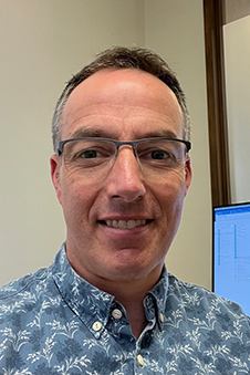
Dr Jon Cornwall.
Dr Cornwall led the development of recently adopted global guidelines for the acquisition and use of images from bodies donated to science.
“The use of images arising from bodies donated to medical science in an expanding digital environment is challenging when many donors have not specifically consented to it,” he says.
When there were only hard copies of images, such as photographs, distribution and appropriate use could be carefully controlled. Now, the increasing use of digital technologies means there is a larger risk of widespread dissemination. Changes in the delivery of education during COVID-19 was a “real eye-opener” in this space because of the big increase in the adoption of digital technology and online sharing of resources.
There were three things that were major considerations when developing the international guidelines, says Dr Cornwall. These were the dignity and respect of donors, what donors consented to and the social mores around how bodies of the deceased should be treated.
Dr Cornwall currently works in medical education and has a research focus on ethical issues surrounding the use of bodies donated to science. He is a member of the Federative International Committee of Ethics and Medical Humanities (FICEM), the group that oversees ethical processes around body donation for the International Federation of Associations of Anatomists (IFAA). He is also one of three academics, together with Tom Champney (University of Miami) and Sabine Hildebrandt (Harvard University), guiding the ‘Bioethics Unicorns’ initiative that seeks to promote the topics of ethics and professionalism within anatomy education.
In 2012, the IFAA published guidelines to support good practice around the use of human bodies and tissues for anatomical purposes. The rise of the internet and other digital technologies means there are now other considerations which require best practice guidance for the anatomy community.
For that reason, the FICEM developed the ‘Recommendations for good practice around human tissue image acquisition and use in anatomy education and research’.
In this context, the term ‘images’ includes photographs and videos of human tissue, as well as images generated by procedures such as ultrasounds and MRI s. The guidelines say while the images are not physical specimens, they are from actual people and deserve special consideration around their acquisition, storage and use.
The use and distribution of images in ways that are not considered ethical can undermine the relationship health and education organisations have with their local communities.
Dr Cornwall says there is limited information on both donors’ and the public’s views around the use of these images, but “what information there is shows people do care about how the images are treated, so those working in this area have to take that into account”.
Kōrero by Andrea Jones, Team Leader, Divisional Communications