An excerpt from He Kitenga Horizons, a University of Otago publication, December 2013
Researchers in Otago's Departments of Physics and Pathology are combining their expertise in two projects that have the potential to revolutionise techniques used for cancer detection and diagnosis.
The potential use of light and ultrasound in cancer detection is being explored by Associate Professor Igor Meglinski and Dr Jevon Longdell (Physics) in association with Professor Michael Eccles (Pathology).
In the first of two collaborations, Meglinski and Longdell aim to develop and demonstrate a new non-invasive method for 3-D biomedical imaging, with an initial application to cancerous tissue.
They are working on a medical device that combines two existing light and ultrasound techniques (multispectral optoacoustic tomography and ultrasound modulated optical tomography) to provide clear, accurate images of tissue, allowing it to be analysed for cancer cells at a much earlier stage than current technology permits and at a fraction of the cost.
Longdell explains that some light is able to travel through the body, but bounces around like a pinball so it cannot be used to make an image. Ultrasound, on the other hand, travels through a person without being scattered very much, but doesn't distinguish between different types of tissue.
By combining the two, they hope to improve on ultrasound imaging by being able to distinguish between normal and abnormal cells on their optical qualities as well.
They are now part way through a two-year first stage of the project during which they are developing a prototype. “We will then have to work more closely with physicians to check the new system and collect enough statistics to demonstrate the equipment is accurate and works for the benefit of patients,” says Meglinksi.
He is confident the combination of light and ultrasound has the potential for other medical uses. “We think it could be used, for example, for vein ulcer analysis or in sports medicine, and there are hopes for its development in brain imaging. The only problem at the moment is the skull, which is difficult to go through. But there are some ideas on how to overcome this problem and, in a few years, this technique might be even better for brain studies than current imaging techniques.”
He adds that, while the research has an initial medical focus, he can envisage wider applications. “I am really pragmatic. This country is very well known for its agricultural products. So we think it might be used, for example, for testing the freshness and healthiness of meat.”
Eccles is providing tissue samples and medical expertise for the project.
In a second collaboration, Meglinski and Eccles are investigating a new way to measure the aggressiveness of cancer cells using a form of laser light (circular polarised light).
Eccles says the research aims to determine whether circular polarised light can be used to identify the grade and aggressiveness of cancer cells due to changes that occur in the optical properties of the cells. When explaining the technique to lay people, he likens it to Dr Leonard “Bones” McCoy in Star Trek waving a device over patients to diagnose their ills and the answer being immediately available on a screen.
Meglinski, who came up with the idea, initially borrowed pathology samples of cancer tissues on slides, put them under circular polarised light and found that he could predict whether they were cancerous.
“We played with the samples for a few months by trying to send light through them. We could see nothing and I said, 'So, okay, my idea is rubbish'. Then we tried to send the light with side reflection and we found that we were able to recognise normal and abnormal cells.”
Eccles says he is particularly interested in whether circular polarised light detects the changes in the nuclei of cancer cells as they become more aggressive and whether this explains the results Meglinski has been achieving.
He says that while the research is at an early stage, it has huge possibilities. “There are a bunch of reasons why it might be better if you can use light. It takes quite an investment in time and effort for a pathologist to get come up with an answer. If it's possible to get the same answer using circular polarised light on a tissue without doing a biopsy you can get the answer quite quickly.
“It could also speed up the whole process so that a patient in a doctor's waiting room might wait only a matter of minutes to have a result. It could then speed up the process for the patient to have surgery if the case is serious. In addition, it might allow a practitioner to assess, in real time, that the cancer is still there after surgery or has been removed.”
FUNDING
Health Research Council, Explorer Grant
Maurice Wilkins Centre
Ministry of Business, Innovation and Employment, Science Investment Round
New Zealand Institute for Cancer Research Trust

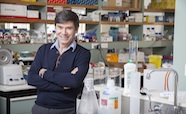
Professor Mike Eccles: "If it's possible to get the same answer (as a pathologist) using circular polarised light on a tissue without doing a biopsy you can get the answer quite quickly"