This page provides a brief description of the electron microscopes and sample preparation equipment.
For more information about techniques visit:
For a more detailed discussion of what the equipment can do, please contact a member of staff for advice.
Electron microscopy team
If you are looking for confocal microscopy equipment please visit:
Confocal microscopy
Registration and training
You are not permitted to use any of the equipment unless you have been trained to a suitable level and understand any health and safety issues associated with each item. You will not be expected to remember everything after one training session; a staff member will be available to assist you at most times. When you are competent at using an item of equipment, you will be allowed after-hours access to the facility.
Specification for publications
This equipment page is useful for finding the name, manufacturer and other specifications when you're writing up a thesis or paper. All company names and addresses are listed as they were at the time of equipment purchase. If you notice something missing which you could have used, please let us know so we can add it.
Microscopes
JEOL 2200FS Cryo-TEM
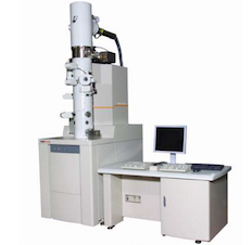 JEOL 2200FS field emission scanning transmission electron microscope (STEM) with omega energy filter (JEOL Ltd, Tokyo, Japan). It is fitted with a TVIPS F416 CMOS camera (TVIPS, Gauting, Germany) and a Direct Electron DE-20 detector (Direct Electron LP, California, U.S.A). HAADF and bright field modes are available for STEM electron detection.
Specimen holders include a standard JEOL holder, a JEOL double-tilt holder and a Gatan model 914 high tilt cryo holder (Gatan, Inc, California, USA) for cryo specimens.
Image acquisition software includes: SerialEM (University of Colorado, Boulder, USA), TVIPS EM-Menu and DE Imaging Manager.
JEOL 6700F FE-SEM
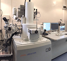 Field emission scanning electron microscope (JEOL Ltd, Tokyo, Japan) fitted with a JEOL 2300F EDS system (JEOL Ltd, Tokyo, Japan) and a Gatan Alto 2500 cryo preparation chamber/cryo stage (Gatan Inc, Pleasanton, California, USA). Our main research scanning electron microscope, capable of preparing and viewing frozen-hydrated specimens and undertaking elemental analysis and mapping.
Zeiss Sigma 300 SBF SEM
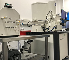 Zeiss Sigma 300 VP FE scanning electron microscope (Carl Zeiss Inc, Oberkocken, Germany) with VPSE-G4 and HDBSD detectors and fitted with Gatan 3ViewXP serial block face system and OnPoint detector (Gatan Inc, Pleasanton, California, USA) and Oxford Ultim 20mm2 Max EDS system with AZtecLive software (Oxford Instruments, Oxfordshire, UK).
This variable pressure SEM system can be used in conventional SEM mode using secondary electron and backscatter imaging for a wide variety of specimens, including uncoated specimens. At the same time it can acquire energy dispersive spectroscopy for composition analysis. Alternatively, when the Gatan 3View2XP serial block face system is fitted, it acquires high resolution volume data using the Gatan OnPoint low kV, high speed BSE detector while specimen charging is controlled by the Zeiss FCC charge compensation system.
Philips CM100 TEM
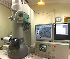 Philips CM100 BioTWIN transmission electron microscope with LaB6 emitter (Philips/FEI Corporation, Eindhoven, Holland). Fitted with MegaView lll digital camera (Olympus Soft Imaging Solutions GmbH, Münster, Germany).
A high-contrast transmission electron microscope designed for biological specimens.
Zeiss Sigma VP FEG SEM
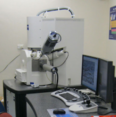 Zeiss Sigma VP variable-pressure scanning electron microscope (Carl Zeiss Inc, Oberkocken, Germany) fitted with a HKL INCA Premium Synergy Integrated ED/EBSD system (Oxford Instruments, Oxfordshire, UK) and modified Emitech K1250 cryo stage (Emitech, UK).
The Zeiss Sigma is able to perform high resolution imaging with secondary and back scatter electrons (up to 1.7nm at 15 kV and 3.0nm at 1 kV). It is able to operate under variable pressure conditions (from 2 to 133 Pa) and is good for imaging specimens that are poorly conductive or specimens that cannot be imaged under vacuum. The chamber is able to accommodate specimens up to 250mm in diameter and 145mm tall.
Sample preparation equipment
Ultramicrotomes
- Leica UC6 Ultramicrotome (Leica Microsystems GmbH Ernst-Leitz-Straße 17–37 Wetzlar, 35578 Germany)
- Leica UC7 Ultramicrotome (Leica Microsystems GmbH Ernst-Leitz-Straße 17–37 Wetzlar, 35578 Germany)
- Leica UCT + FCS cryo ultramicrotome (Leica Microsystems GmbH Ernst-Leitz-Straße 17–37 Wetzlar, 35578 Germany)
Vacuum coating systems
- Quorum Q150T ES coater (Quorum Technologies, East Sussex, UK) for carbon coating
- Quorum Q150V ES PLUS coater (Quorum Technologies, East Sussex, UK) for sputter coating (Au/Pd, etc)
- EMS/Quorum GloQube glow discharge system (Quorum Technologies, East Sussex, UK) for surface charge modification of grids, etc
- Edwards E306A vacuum coating system (Edwards High Vacuum, Crawley, England) for special coating tasks, including double coating etc
Conventional sample preparation equipment
- Lynx el Tissue Processor (Australian Biomedical Corporation Ltd, Mount Waverley, Vic. 3149, Australia) for automated fixation, dehydration, staining and resin infiltration of specimens.
- Leica EM AC20 (Leica Microsystems GmbH Ernst-Leitz-Straße 17-37 Wetzlar, 35578 Germany) for contrasting ultra-thin sections on grids
- LKB 2168 Ultrostain grid stainer (LKB-Produkter AB, Bromma, Sweden) for contrasting ultra-thin sections on grids
- Leica EM IGL automatic immunolabeling device (Leica Microsystems, Vienna, Austria) for automated immunolabelling of grid-mounted electron microscopy specimens
- Pelco BioWave Pro laboratory microwave oven with ColdSpot heating reduction system (Ted Pella Inc, Redding, California, USA) for microwave-stimulated decalcification, fixing, dehydration and infiltration of specimens.
- Eppendorf 5810 centrifuge (Eppendorf AG, Hamburg, Germany)
- Heraeus Sepatech Biofuge 15 centrifuge (Heraeus Sepatech GmbH, Osterode, Germany)
- Wescor Vapro 5600 osmometer (ELITech Group Inc, South Logan, Utah, USA)
Cryo and hybrid sample preparation equipment
- Leica EM Pact2 high pressure freezing device (Leica Microsystems GmbH, Vienna, Austria) for rapid specimen freezing
- Vitrobot Mk IV cryo-TEM specimen freezing system (FEI Co. Hillsboro, Oregon Thermo Fisher Scientific, USA) for rapid specimen freezing on grids
- Reichert KF80 specimen plunge-freezing device with metal mirror (MM80) attachment (C. Reichert Optische Werke AG, Vienna, Austria) for rapid specimen freezing
- Reichert/Leica AFS automatic freeze-substitution device (Reichert Division of Leica AG, Vienna, Austria) and;
Leica EMAFS2 automatic freeze-substitution device (Leica Microsystems GmbH, Vienna, Austria)
– both for freeze-substitution of frozen specimens, for the progressive lowering of temperature technique for chemically-fixed specimens and low-temperature resin curing - Bal-Tec BAF 060 freeze fracture device (Bal-Tec AG, Balzers, Liechtenstein) for replica creation and shadow contrasting of frozen and fractured specimens
- Bal-Tec CPD-030 critical point dryer (Bal-Tec AG, Balzers, Liechtenstein) for drying of solvent-dehydrated specimens